Articles by "disease"

Showing posts with label disease. Show all posts

are
Uterine fibroids

Uterine fibroids (also called leiomyomas or myomas) are benign growths of the muscle inside the uterus. They are not cancerous, nor are they related to cancer. Fibroids can cause a wide variety of symptoms, including heavy menstrual bleeding and pressure on the pelvis.

Description

Uterine fibroids are extremely common. About 25% of women in their reproductive years have noticeable fibroids. There are probably many more women who have tiny fibroids that are undetected.

Fibroids develop in women between the ages of 30–50. They are never seen in women younger than 20 years old. After menopause, if a woman does not take estrogen, fibroids shrink. It appears that African American women are much more likely to develop uterine fibroids.

areare

Fibroids are divided into different types, depending on the location. Submucous fibroids are found in the uterine cavity; intramural fibroids grow on the wall of the uterus; and subserous fibroids are located on the outside of the uterus. Many fibroids are so large that they fit into more than one category. The symptoms caused by fibroids are often related to their location.

Causes and symptoms

No one knows exactly what causes fibroids. The growth of fibroids, however, appears to depend on the hormone estrogen. Fibroids often grow larger when estrogen levels are high, as in pregnancy. Medications that lower the estrogen level can cause the fibroids to shrink.

The signs and symptoms of fibroids include:
  • Heavy uterine bleeding. This is the most common symptom, occurring in 30% of women who have fibroids. The excess bleeding usually happens during the menstrual period. Flow may be heavier, and periods may last longer. Women who have submucous or intramural fibroids are most likely to have heavy uterine bleeding.
  • Pelvic pressure and pain. Large fibroids that press on nearby structures such as the bladder and bowel can cause pressure and pain. Larger fibroids tend to cause more severe symptoms.
  • Infertility. This is a rare symptom of fibroids. It probably accounts for less than 3% of infertility cases. Fibroids can cause infertility by compressing the uterine cavity. Submucous fibroids can fill the uterine cavity and interfere with implantation of the fertilized egg.
  • Miscarriage. This is also an unusual symptom of fibroids, probably accounting for only a tiny fraction of the miscarriages that occur.
  • Pregnancy complications. Fibroids can greatly increase in size during pregnancy, because of increased levels of estrogen. They can cause pain, and even lead to premature labor.

Diagnosis

A healthcare provider can usually feel fibroids during a routine pelvic examination. Ultrasound can be used to confirm the diagnosis, but this is not generally necessary.

Treatment

There are several natural treatments that help lower estrogen levels and slow the growth of the benign tumors. One study of alternative and complementary treatments for uterine fibroids found, however, that the cost of the alternative remedies was significantly higher than the cost of conventional treatments.

Nutritional therapy

There are several things women can do nutritional-wise to avoid having fibroids or prevent them from getting bigger:
  • Eat more fruits, green or sea vegetables, whole grains, nuts, and seeds.
  • Eat more soy foods such as tofu, tempeh, miso, or soy burger. Soy products contain isoflavones, which help reduce high levels of estrogens in the body.
  • Avoid foods with high fat or sugar content, caffeine,or alcohol.
  • Avoid eating produce sprayed with insecticides.

Nutritional supplements

The following supplements may be helpful in lowering estrogen levels and controlling fibroids:
  • Bromelain: reduces inflammation.
  • Choline: may improve liver function.
  • Flaxseed: helps reduce excessive production of estrogens and other hormones.
  • Vitamin E and evening primrose oil: help to regulate hormone production and may even shrink the fibroids.
  • Vitamin C and bioflavonoids: have antiinflammatory and antioxidant effects.

Herbal treatment

Kuei-chih-fu-ling-wan (Keishi-bukuryo-gan; KBG) is a traditional Chinese herbal preparation that can effectively shrink fibroid tumors in 60% of patients, according to one study conducted by Japanese scientists. KBG is a mixture of the following herbs: cassia bark (Keihi), herbaceous peony roots (Shakuyaku), peach kernels (Tounin), herbaceous fungus (Bukuryo), and root bark of peony (Botanpi).

In addition to reducing fibroid size, KBG also successfully alleviated fibroid symptoms such as severe menstrual bleedings or menstrual pain in 90% of the women in the study. These researchers suggest that KBG may work by inhibiting the production of sex hormones including estrogen.

Unlike many other presently available herbal preparations that may be effective but lack scientific evidence to support their uses, KBG is proven safe as well as having few side effects. Women with fibroids, therefore, have one more alternative treatment to hysterectomy.

There are many herbal formulas that can be used depending on specific symptoms and body types. Another herbal treatment that may also be effective is wild yam progesterone cream. However, these are potent drugs and patients should consult their doctors before trying any of these treatments.

Homeopathy

A homeopathic physician may prescribe patient-specific homeopathic remedies to control fibroid symptoms.

Allopathic treatment

Not all fibroids cause symptoms. Even fibroids that do cause symptoms may not require treatment. In the majority of cases, the symptoms are inconvenient and unpleasant, but do not result in health problems.

Occasionally, fibroids lead to such heavy menstrual bleeding that the woman becomes severely anemic. In these cases, treatment of the fibroids may be necessary. Very large fibroids are much harder to treat. Therefore, many doctors recommend treatment for moderatelysized fibroids, in order to prevent them from growing into large fibroids that cause worse symptoms.

The following are possible treatment plans:
  • Observation (watchful waiting). Most women already have symptoms at the time their fibroids are discovered, but feel that they can tolerate their symptoms. Therefore, no active treatment is given, but the woman and her physician stay alert for signs that the condition might be getting worse.
  • Hysterectomy. This procedure involves surgical removal of the uterus, and it is the only definitive cure for fibroids. In fact, 25% of hysterectomies are performed because of symptomatic fibroids. A gynecologist can remove a fibroid uterus during either an abdominal or a vaginal hysterectomy. The choice depends on the size of the fibroids and other factors such as previous births and previous surgeries.
  • Myomectomy. In this surgical procedure only the fibroids are removed; the uterus is repaired and left in place. This is the surgical procedure many women choose if they are not finished with childbearing. At first glance, it seems that this treatment is a middle ground between observation and hysterectomy. However, myomectomy is actually a difficult surgical procedure, more difficult than a hysterectomy. Myomectomy often causes significant blood loss, and blood transfusions may be required. In addition, some fibroids are so large, or buried so deeply within the wall of the uterus, that it is not possible to save the uterus, and a hysterectomy must be done, even though it was not planned.
  • Lowering estrogen levels. Since fibroids are dependent on estrogen for their growth, medical treatments that lower estrogen levels can cause fibroids to shrink. A group of medications known as GnRH antagonists can dramatically lower estrogen levels. Women who take these medications for three to six months find that their fibroids shrink in size by 50% or more. They usually experience dramatic relief of their symptoms of heavy bleeding and pelvic pain.
  • Uterine artery embolization (UAE). Embolization is a newer alternative to hysterectomy that shrinks fibroids by cutting off their blood supply. In UAE, the surgeon inserts a catheter into the uterine arteries. Small particles of polyvinyl foam or other inert substances are injected through the catheter into the arteries. The particles form an embolus, or clump, that blocks the blood supply to the fibroids and causes them to shrink. UAE is still controversial, however, because it has been associated with significant complications.

Unfortunately, GnRH antagonists cause unpleasant side effects in over 90% of women. The therapy is usually used for only three months, and should not be used for more than six months because the risk of developing brittle bones (osteoporosis) begins to rise.

Once the treatment is stopped, the fibroids begin to grow back to their original size. Within six months, most of the old symptoms return. Therefore, GnRH agonists cannot be used as long-term solution.
are
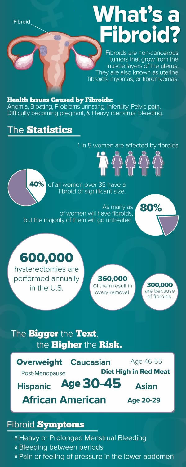
Uterine fibroids infographic

At the moment, treatment with GnRH antagonists is used mainly in preparation for surgery (myomectomy or hysterectomy). Shrinking the size of the fibroids makes surgery much easier, and reducing the heavy bleeding allows a woman to build up her blood count before surgery.

Fibroids can cause problems during pregnancy because they often grow in size. Large fibroids can cause pain and lead to premature labor.

Fibroids cannot be removed during pregnancy because of the risk of injury to the uterus and hemorrhage. GnRH antagonists cannot be used during pregnancy. Treatment is limited to pain medication and medication to prevent premature labor, if necessary.

Expected results

Many women who have fibroids have no symptoms or have only minor symptoms of heavy menstrual bleeding or pelvic pressure. However, fibroids tend to grow over time, and gradually cause more symptoms. Many women ultimately decide to have some form of treatment. Currently, hysterectomy is the most popular form of treatment.

Prevention

Eating a healthful diet, reducing stress, and exercising regularly is the preferred preventive treatment regimen of many diseases including fibroids.

Uterine
Uterine cancer

Uterine cancer can be divided into two primary forms, cervical and endometrial. Cancer of the cervix most often affects the neck of the cervix or the opening or the opening into the uterus from the vagina. Endometrial cancer affects the inside lining of the uterus.

Cervical cancer is much more prevalent than cancer of the endometrium; some estimate the incidence ratio as 3:1. Statistics from the year 2000 indicated cervical cancer was the second leading cause of cancer deaths in women ages 20-39 years, and the fifth leading cause of cancer

death in women from 40-59 years old. Unlike many other cancers, early cancer of the cervix can be identified as much as 10 or more years before the cancer invades other tissues. These visible changes in the structure and activity of the cervical cells are seen under the microscope with Papanicolaou (Pap) testsUterine and are referred to as mild dysplasia.

Uterine Uterine

Over a time period of five to 10 years, these aneh cells may disappear without treatment, or may invade into deeper tissues and progress into a true cancer. The cancerous cells then may spread to endometrium, lymph glands, and nerves in the pelvic region.

As the population ages, cancer of the endometrium is becoming more common. Statistics indicate that approximately 50% of women with postmenopausal bleeding are diagnosed with endometrial cancerUterine.

This early symptom of irregular vaginal bleeding often allows removal of the uterus to result in cure of the disease, as endometrial cancer progresses and spreads slowly.

While all women are at risk for developing uterine cancer, specific risk factors for cervical cancer include sexual activity at an early age, and sex with multiple partners.

Infertility, diabetes, obesity, and estrogen therapy place a woman at high risk for endometrial cancer. Other risk factors for uterine cancer include: endometrial hyperplasia, sexual inactivity, undergoing menopause after age 59 years, and never having had children.

Causes and symptoms

Uterine
A completed cancer awareness ribbon for sarcoma
use pink for breast cancer, or peach for uterine cancer.

An important factor linked to cervical cancer is infection with one of the most common sexually transmitted diseases—warts while others have no observable symptoms.

Individuals infected with the herpes simplex virusUterine, human immunodeficiency virus (HIV) or acquired immune deficiency syndrome (AIDS) are at increased risk for developing cancer of the cervix; the associated suppression of the immune system allows the HPV to more easily invade. Other chronic infections and erosions of the cervix also may increase the risk of cervical cancer.

While some women who have precancerous cervical changes experience no symptoms, others notice heavier or longer menstrual periods or vaginal bleeding after douching, intercourse or between periods.

Symptoms of more advances stages of uterine cancer may include a foul-smelling vaginal discharge, rectal pressure or constipation, loss of appetite, fatigue, and back or leg pain.

Diagnosis

An annual Pap test and pelvic examination beginning as soon as young woman becomes sexually active, or between the age 17-20 years, are the most important diagnostic steps for early detection of uterine cancer.

The Pap smear can pick up cervical dyplasia and the conventional physican may then perform a colposcopy and biopsy of the cervix to give a better understanding of the abnormalities.

If only a small area of the cervix is affected, the recommendation may be made for more frequent Pap tests (about every three to six months) to monitor for changes in the cells of the cervix. Additional diagnostic tests for uterine cancer may include laparoscopy, laparotomy, or vaginosonography.

In 2002, a Food and Drug Administration (FDA) advisory panel suggested adding a screening test for HPV in addition to Pap smears since HPV is such a high risk factor for cervical cancer. The screening test could help separate women at high risk for more frequent screening than women not at high risk.

Treatment

pinky uterus

After cervical dysplasia has been found, several herbal remedies and supplements may be helpful. Practitioners of herbal medicine refer to this class of herbs as emmenagogues.

It includes supplements such as selenium, and vitamins B6 and C.

While these studies make no claim that taking a multivitamin or mineral supplement can reverse advanced cervical dysplasia, taking these supplements preventively may make sense.

The woman with uterine cancer will also benefit from nutritional supplements and a diet aimed at strengthening the immune system. EchinaceaUterine and garlic supplementsUterine may not only have positive effects on immunities, but also counteract the side effects of cancer treatment.

Many trace elements, flavonoids, and other phytochemicals are provided by eating a well balanced diet that may not be provided in a pill. Even with relatively low levels of dietary intake, shiitake mushrooms, lentinus edodesUterine, laminaria sea vegetables, and kombu kelpUterine are believed to have anticancer properties.

The use of any supplements or specific dietary modification should be discussed with the physician treating the cancer in order to avoid any undesirable drug interactions or side effects.

Research emphatically supports the mind-body connection when considering the health of the individual with cancer. Studies have also shown the positive effects of imagery on boosting immunities and natural killer cells.

Visualization of the dominant white blood cells successfully attacking weak cancer cells can not only have a positive effect on the mood and mental status, but may also shrink tumors and extend the life of a patient with cancer. Laughter has also been found to enhance immunities and stimulate the sympathetic nervous system, pituitary gland, and the hormones that reduce stress, inflammation, and pain.

In addition to the well known effects of massage for relaxation and stress reduction, there are other physiologic effects that may help the individual with cancer.

Massage may slow the body’s release of the stress hormone cortisol, decreasing anxiety and allowing for more effective periods of sleep and regeneration. Massage has also been found to increase the production of serotonin, which can improve overall mood and immune status.

Allopathic treatment

Early stages of cervical dysplasia may require only frequent reevaluation to monitor progression or regression of the aneh cells. Regression of aneh cells may occur due to the immune response or lifestyle changes, such as discontinuing smoking or oral contraceptive use. In more advanced cases, the cervical lining may be removed via cautery, freezing or laser procedures.

Age, overall health status, and the presence of other aneh findings will impact on the selection of most appropriate treatment plan for uterine cancer. Surgery may be presented as a treatment option for invasive cancer.

Extent of the surgical procedures will depend upon the stage of the cancer. A hysterectomy, lymphadenectomy, or total pelvic exenteration may be recommended. Radiation therapyUterine may be offered instead of or in addition to surgical removal of the affected tissues.

Depending on the individual’s disease stage, and the response and tolerance to the radiation, treatment may be provided by external beams directed over the pelvis, or by the insertion of radium tubes into the uterus and/or vagina. Chemotherapy may also be recommended, involving the infusion of tumor-fighting drugs directly into the circulatory system.

Expected results

The outcomes for the individual with uterine cancer are significantly related to the stage of the disease when cancer is found and treatment initiated. Early interventions can result in nearly 100% cure rates, while those individuals whose cancer is not discovered until aneh tissue growth has invaded surrounding organs may have less positive outcomes. Those with advanced disease may experience pain, vaginal bleeding and/or foul smelling discharge, and intestinal obstruction.

Prevention

The best preventive measure against uterine cancer is an annual pelvic examination and Pap test. In fact, a 2002 report from the College of American Pathologists stated that 80 percent of the women who die from cervical cancer had not had a Pap test in the five years preceding their diagnosis.

Recognition of risk factors for uterine cancer, along with an awareness of the early signs and symptoms of cervical dysplasia, can promote the early detection of changes in the cervical cells.

An
Digestive Ulcers

An ulcer is an eroded area of skin or mucous membrane. In common usage, however, ulcer usually refers to disorders in the upper digestive tract. The terms ulcer, gastric ulcer, and peptic ulcer are often used interchangeably.

Peptic ulcers can develop in the lower part of the esophagus, the stomach, the first part of the small intestine (the duodenum), and the second part of the small intestine (the jejunum).

It is estimated that 2% of the adult population in the United States has active digestive ulcers, and that about 10% will develop ulcers at some point in their lives. There are about 500,000 new cases in the United States every year, with as many as 4 million recurrences. The male/female ratio for digestive ulcers is 3:1.

AnAn

The most common forms of digestive ulcer are duodenal and gastric. About 80% of all digestive ulcers are duodenal ulcers. This type of ulcer may strike people in any age group but is most common in males between the ages of 20 and 45. The incidence of duodenal ulcers has dropped over the past 30 years. Gastric ulcers account for about 16% of digestive ulcers.

They are most common in males between the ages of 55 and 70. The most common cause of gastric ulcers is the use of nonsteroidal anti-inflammatory drugs, or NSAIDs. The current widespread use of NSAIDs is thought to explain why the incidence of gastric ulcers in the United States is rising.

Causes and symptoms

Causes of ulcers

There are three major causes of digestive ulcers: infection; certain medications; and disorders that cause oversecretion of stomach juices.

HELICOBACTER PYLORI INFECTION. Helicobacter pylori is a bacterium that lives in the mucous tissues that line the digestive tract. Infection with H. pylori is the most common cause of duodenal ulcers. About 95% of patients with duodenal ulcers are infected with H. pylori, as opposed to only 70% of patients with gastric ulcers. USE OF NONSTEROIDAL ANTI-INFLAMMATORY

DRUGS (NSAIDS). Nonsteroidal anti-inflammatory drugs, or NSAIDs, are painkillers that many people use for headaches, sore muscles, arthritis, and menstrual cramps. Many NSAIDs are available without prescriptions. Common NSAIDs include aspirin, ibuprofen (Advil, Motrin), flurbiprofen (Ansaid, Ocufen), ketoprofen (Orudis), and indomethacin (Indacin).

Chronic NSAID users have 40 times the risk of developing a gastric ulcer as nonusers. Users are also three times more likely than nonusers to develop bleeding or fatal complications of ulcers. Aspirin is the most likely NSAID to cause ulcers.

Other Risk Factors
  • Hypersecretory syndromes, including Zollinger-Ellison syndrome, secrete excessive amounts of digestive juices into the digestive tract. Fewer than 5% of digestive ulcers are due to these disorders.
  • Smoking increases a patient’s chance of developing an ulcer, decreases the body’s response to therapy, and increases the chances of dying from complications.
  • Blood type. Persons with type A blood are more likely to have gastric ulcers, while those with type O are more likely to develop duodenal ulcers.
  • Attitudes toward stress, rather than the presence of stress, puts one at risk for ulcers.
  • Having a critical illness. Patients who are very sick are at increased risk of developing stress-related ulcers.
The consumption of high-fat or spicy foods is not a significant risk factor.

Symptoms

Not all digestive ulcers produce symptoms; as many as 20% of ulcer patients have so-called painless or silent ulcers. Silent ulcers occur most frequently in the elderly and in chronic NSAID users.

The symptoms of gastric ulcers include feelings of indigestion and heartburn, weight loss, and repeated episodes of gastrointestinal bleeding. Ulcer pain is often described as gnawing, dull, aching, or resembling hunger pangs. The patient may be nauseated and suffer loss of appetite.

About 30% of patients with gastric ulcers are awakened by pain at night. Many patients have periods of chronic ulcer pain alternating with symptom-free periods that last for several weeks or months. This characteristic is called periodicity.

The symptoms of duodenal ulcers include heartburn, stomach pain relieved by eating or taking antacids, weight gain, and a burning sensation at the back of the throat. The patient is most likely to feel discomfort two to four hours after meals, or after having citrus juice, coffee, or aspirin.

About 50% of patients with duodenal ulcers awake during the night with pain, usually between midnight and 3 A.M. A regular pattern of ulcer pain associated with certain periods of day or night or a time interval after meals is called rhythmicity.

An
Foods that make ulcers worse

Complications

Between 10%–20% of peptic ulcer patients develop complications at some time during the course of their illness. All of these are potentially serious conditions. Complications are not always preceded by diagnosis of or treatment for ulcers; as many as 60% of patients with complications have not had prior symptoms.

Bleeding is the most common complication of ulcers. It may result in anemia, vomiting blood, or the passage of bright red blood through the rectum. The mortality rate from ulcer hemorrhage is 6-10%.

About 5% of ulcer patients develop perforations, which are holes through which the stomach contents can leak out into the abdominal cavity. The incidence of perforation is rising because of the increased use of NSAIDs, particularly among the elderly.

The signs of an ulcer perforation are severe pain, fever, and tenderness when the doctor touches the abdomen. Most cases of perforation require emergency surgery. The mortality rate is about 5%.

Ulcer penetration is a complication in which the ulcer erodes through the intestinal wall without digestive fluid leaking into the abdomen. Instead, the ulcer penetrates into an adjoining organ, such as the pancreas or liver. The signs of penetration are more severe pain without rhythmicity or periodicity, and the spread of the pain to the lower back.

Obstruction of the stomach outlet occurs in about 2% of ulcer patients. It is caused by swelling or scar tissue formation that narrows the opening between the stomach and the duodenum (the pylorus). Over 90% of patients with obstruction have recurrent vomiting of partly digested or undigested food; 20% are seriously dehydrated.

Diagnosis

Physical examination and patient history

The diagnosis of peptic ulcers is rarely made on the basis of a physical examination alone. The only significant finding may be mild soreness in the area over the stomach when the doctor presses (palpates) it. The doctor is more likely to suspect an ulcer if the patient has one or more of the following risk factors:
  • member of the male sex
  • age over 45
  • recent weight loss, bleeding, recurrent vomiting, jaundice, back pain, or anemia
  • history of using aspirin or other NSAIDs
  • history of heavy smoking
  • family history of ulcers or stomach cancer

Endoscopy and imaging studies

An endoscopy is considered the best procedure for diagnosing ulcers and taking tissue samples. An endoscope is a slender tube-shaped instrument used to view the tissues lining the stomach and duodenum. If the ulcer is in the stomach, then a tissue sample will be taken because 3-5% of gastric ulcers are cancerous.

Duodenal ulcers are rarely cancerous. Radiological studies are sometimes used instead of endoscopy because they are less expensive, more comfortable for the patient, and are 85% accurate in detecting cancer.

Laboratory tests

Blood tests usually give normal results in ulcer patients without complications. They are useful, however, in evaluating anemia from a bleeding ulcer or a high white cell count from perforation or penetration. Serum gastrin levels can be used to screen for Zollinger-Ellison syndrome.

It is important to test for H. pylori because almost all ulcer patients who are not taking NSAIDs are infected. Noninvasive tests include blood tests for immune response and a breath test. In the breath test, the patient is given an oral dose of radiolabeled urea.

If H. pylori is present, it will react with the urea and the patient will exhale radiolabeled carbon dioxide. Invasive tests for H. pylori include tissue biopsies and cultures performed from fluid obtained by endoscopy.

Treatment

Alternative treatments can relieve symptoms and promote healing of ulcers. A primary goal of these treatments is to rebalance the stomach’s hydrochloric acid output and to enhance the mucosal lining of the stomach.

Food allergies have been considered a major cause of stomach ulcers. An elimination/challenge diet can help identify the allergenic food(s) and continued elimination of these foods can assist in healing the ulcer.

Ulcer patients should avoid aspirin, stop smoking, avoid antacids, and reduce stress. Dietary changes include avoidance of sugar, caffeine, and alcohol, and reducing milk intake.

Supplements

Dietary supplements that help to control ulcer symptoms include:

Herbals

Botanical medicine offers the following remedies that may help treat ulcers:

Chinese medicines

Chinese herbal treatment principles are based upon specific groups of symptoms. Chinese patent medicines are also based upon specific symptoms and include:
  • Wu Bei San (cuttlefish bone and fritillaria): acid reflux and bleeding
  • Wu Shao San (cuttlefish bone and paeonia): acid reflux and bleeding
  • Liang Fu Wan (galangal and cyperus pill): pain
  • 204 Wei Tong Pian (204 epigastric pain tablet): pain, acid reflux, and bleeding
  • Xi Lei San (tin-like powder): ulcer with tarry stool

Other treatments

Other treatments for ulcers are:
  • Essence therapy. Dandelion essence can help reduce tension, and pink yarrow essence can help the patient distinguish between his or her problems and those of others.
  • Reflexology. For ulcers, the practitioner work the solar plexus and stomach points on the feet and the solar plexus, stomach, and top of shoulder points on the hands.
  • Biofeedback. Thermal biofeedback can help protect and heal the stomach.
  • Sound therapy. Music with a slow, steady beat can promote relaxation and reduce stress.
  • Ayurveda. Ayurvedic treatment is individualized to each patient but common ulcer remedies include: aloe vera natural gel, arrowroot powder with hot milk, and tea prepared from cumin, coriander, and fennel seeds.
  • Acupuncture. Ulcers can be treated using target points for stress, anxiety, and stomach problems.
  • Relaxation techniques. Stress reduction and involvement in stress management programs may help relieve ulcer symptoms.

Allopathic treatment

Medications

Most drugs that are used to treat ulcers work by either lowering the rate of stomach acid secretion or protecting the mucous tissues that line the digestive tract.

Medications that lower the rate of stomach acid secretions fall into two major categories: proton pump inhibitors and H2 receptor antagonists.

The proton pump inhibitors, which have been in use since the early 1990s, include omeprazole (Prilosec) and lansoprazole (Prevacid). The H2 receptor antagonists include ranitidine (Zantac), cimetidine (Tagamet), famotidine (Pepcid), and nizatidine (Axid).

Drugs that protect the stomach tissues are sucralfate (Carafate), bismuth preparations, and misoprostol (Cytotec).

Most doctors presently recommend treatment to eliminate H. pylori to prevent ulcer recurrences. Without such treatment, ulcers recur at the rate of 80% per year.

The drug combination used to eliminate the bacterium is tetracycline, bismuth subsalicylate (Pepto-Bismol), and metronidazole (Metizol). Eradication is not always successful, however, for reasons that are unclear.

Surgery

Surgical treatment of ulcers is generally used only for complications and suspected cancer. The introduction of a newer technique for repairing perforated ulcers using a laparoscope rather than opening the patient’s abdomen may reduce some of the risks associated with surgical treatment of ulcers.

The most common surgical procedures are vagotomies, in which the connections of the vagus nerve to the stomach are cut to reduce acid secretion; and antrectomies, which involve the removal of part of the stomach.

Expected results

The prognosis for recovery from ulcers is good for most patients. Very few ulcers fail to respond to the medications that are currently used to treat them. Recurrences can be cut to 5% by eradication of H. pylori. Most patients who develop complications recover without problems even when emergency surgery is necessary.

Prevention

Strategies for the prevention of ulcers or their recurrence include the following:
  • giving misoprostol to patients who must take NSAIDs
  • participating in integrated stress management programs
  • avoiding unnecessary use of aspirin and NSAIDs
  • improving the nutritional status of critically ill patients
  • quitting smoking
  • cutting down on alcohol, tea, coffee, and sodas containing caffeine
  • eating high-fiber foods

is
Tuberculosis

Tuberculosis (TB) is a contagious and potentially fatal disease that can affect almost any part of the body but manifests mainly as an infection of the lungs. It is caused by a bacterial microorganism, the tubercle bacillus or Mycobacterium tuberculosis. TB infection can either be acute and short-lived or chronic and long-term.

Although TB can be prevented, treated, and cured with proper treatment and medications, scientists have never been able to eliminate it entirely. The organism that causes tuberculosis, popularly known as consumption, was discovered in 1882.

Because antibiotics were unknown, the only means of controlling the spread of infection was to isolate patients in private sanatoria or hospitals limited to patients with TB—a practice that continues to this day in many countries. TB spread very quickly and was a leading cause of death in Europe.

isis

At the turn of the twentieth century more than 80% of the people in the United States were infected before age 20, and tuberculosis was the single most common cause of death. Streptomycin was developed in the early 1940s and was the first antibiotic effective against the disease.

The number of cases declined until the mid- to late-1980s, when overcrowding, homelessness, immigration, decline in public health inspections, decline in funding, and the AIDS epidemic caused a slight resurgence of the disease. The increase in TB in the United States peaked in 1992, and new cases reported in the United States continue to decrease as of 2004.

Yet the number of cases in foreign-born individuals is rising, and the number of deaths from TB has been rising, making TB a leading cause of death from infection throughout the world. It is estimated that in the next 10 years 90 million new cases of TB will be reported, with the result of 30 million deaths, or about 3 million deaths per year.

Several demographic groups are at a higher risk of contracting tuberculosis. Tuberculosis is more common in elderly persons. More than one-fourth of the nearly 23,000 cases of TB in the United States in 1995 were reported in people above age 65. TB also is more common in populations where people live under conditions that promote infection, such as homelessness and injection drug use.

In the late 1990s, two-thirds of all cases of TB in the United States affected African Americans, Hispanics, Asians, and persons from the Pacific Islands. Finally, the high risk of TB includes people who have a depressed immune system.

High-risk groups include alcoholics, people suffering from malnutrition, diabetics, and AIDS patients — and those infected by human immunodeficiency virus (HIV) — who have not yet developed clinical signs of AIDS. TB is the number one killer of women of childbearing age worldwide. In poor countries, women with TB often don’t know they have the disease until symptoms become severe.

As of late 2002, TB is a major health dilema in certain immigrant communities, such as the Vietnamese in southern California. One team of public health experts in North Carolina maintains that treatment for tuberculosis is the most pressing healthcare need of recent immigrants to the United States.

In some cases, the vulnerability of immigrants to tuberculosis is increased by occupational exposure, as a recent outbreak of TB among Mexican poultry farm workers in Delaware indicates. Other public health experts are recommending tuberculosis screening at the primary care level for all new immigrants and refugees.

is
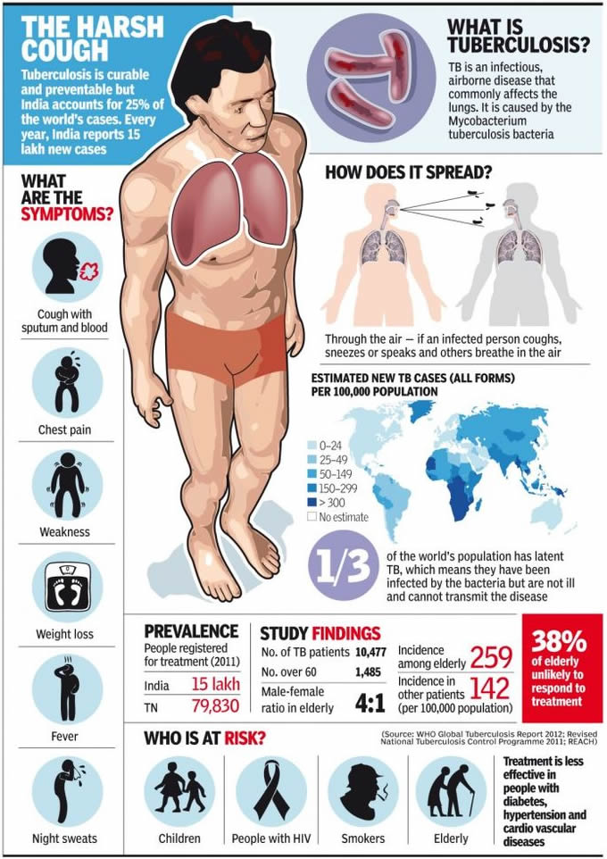
Tuberculosis infographic

Causes and symptoms

Transmission

Tuberculosis spreads by droplet infection, in which a person breathes in the bacilli released into the air when a TB patient exhales, coughs, or sneezes.

However, TB is not considered highly contagious compared to other infectious diseases. Only about one in three people who have close contact with a TB patient, and fewer than 15% of more remote contacts, are likely to become infected.

Unlike many other infections, TB is not passed on by contact with a patient’s clothing, bed linens, or dishes and cooking utensils. Yet if a woman is pregnant, her fetus may contract TB through blood or by inhaling or swallowing the bacilli present in the amniotic fluid.

Once inhaled, water in the droplets evaporates and the tubercle bacilli may reach the small breathing sacs in the lungs (the alveoli), then spread through the lymph vessels to nearby lymph nodes. Sometimes the bacilli move through blood vessels to distant organs.

At this point they may either remain alive but inactive (quiescent), or they may cause active disease. The likelihood of acquiring the disease increases with the concentration of bacilli in the air, and the seriousness of the disease is determined by the number of bacteria with which a patient is infected.

Ninety percent of patients who harbor M. tuberculosis do not develop symptoms or physical evidence of the disease, and their x rays remain negative. They are not contagious; however, these individuals may get sick at a later date and then pass on TB to others.

Though it is impossible to predict whether a person’s disease will become active, researchers surmise that more than 90% of cases of active tuberculosis come from this pool of people. An estimated 5% of infected persons get sick within 12-24 months of being infected.

Another 5% heal initially but, after years or decades, develop active tuberculosis. This form of the disease is called reactivation TB, or post-primary disease. On rare occasions a previously infected person gets sick again after a second exposure to the tubercle bacillus.

Pulmonary tuberculosis

Pulmonary tuberculosis is TB that affects the lungs, and represents about 85% of new cases diagnosed. It usually presents with a cough, which may or may not produce sputum. In time, more sputum is produced that is streaked with blood.

The cough may be present for weeks or months and may be accompanied by chest pain and shortness of breath. Persons with pulmonary TB often run a low-grade fever and suffer from night-sweats. The patient often loses interest in food and may lose weight.

If the infection allows air to escape from the lungs into the chest cavity (pneumothorax) or if fluid collects in the pleural space (pleural effusion), the patient may have difficulty breathing. The TB bacilli may travel from the lungs to lymph nodes in the sides and back of the neck. Infection in these areas can break through the skin and discharge pus.

Extrapulmonary tuberculosis

Although the lungs are the major site of damage caused by tuberculosis, many other organs and tissues in the body may be affected. About 15% of newly diagnosed cases of TB are extrapulmonary, with a higher proportion of these being HIV-infected persons. The usual progression of the disease is to begin in the lungs and spread to locations outside the lungs (extrapulmonary sites).

In some cases, however, the first sign of disease appears outside the lungs. The many tissues or organs that tuberculosis may affect include:
  • Bones. TB is particularly likely to attack the spine and the ends of the long bones.
  • Kidneys. Along with the bones, the kidneys are probably the most common site of extrapulmonary TB. There may, however, be few symptoms even though part of a kidney is destroyed.
  • Female reproductive organs. The ovaries in women may be infected; TB can spread from them to the peritoneum, which is the membrane lining the abdominal cavity.
  • Abdominal cavity. Tuberculous peritonitis may cause pain ranging from the mild discomfort of stomach cramps to intense pain that may mimic the symptoms of appendicitis.
  • Joints. Tubercular infection of joints causes a form of arthritis that most often affects the hips and knees.
  • Meninges. The meninges are tissues that cover the brain and the spinal cord. Infection of the meninges by the TB bacillus causes tuberculous meningitis, a condition that is most common in young children and the elderly. It is extremely dangerous. Patients develop headaches, become drowsy, and eventually comatose. Permanent brain damage can result without prompt treatment.
  • Skin, intestines, adrenal glands, and blood vessels. All these parts of the body can be infected by M. tuberculosis. Infection of the wall of the body’s main artery (the aorta), can cause it to rupture with catastrophic results. Tuberculous pericarditis occurs when the membrane surrounding the heart (the pericardium) is infected and fills up with fluid that interferes with the heart’s ability to pump blood.
  • Miliary tuberculosis. Miliary TB is a life-threatening condition that occurs when large numbers of tubercle bacilli spread throughout the body. Huge numbers of tiny tubercular lesions develop that cause marked weakness and weight loss, severe anemia, and gradual wasting of the body.

Diagnosis

TB is diagnosed through laboratory test results. The standard test for tuberculosis infection, the tuberculin skin test, detects the presence of infection, not of active TB. Skin testing has been done for more than 100 years. In this process, tuberculin is an extract prepared from cultures of M. tuberculosis.

It contains substances belonging to the bacillus (antigens) to which an infected person has been sensitized. When tuberculin is injected into the skin of an infected person, the area around the injection becomes hard, swollen, and red within one to three days.

Today skin tests utilize a substance called purified protein derivative (PPD) that has a standard chemical composition and is therefore a good measure of the presence of tubercular infection.

The PPD test, also called the Mantoux test, is not always 100% accurate; it can produce false positive as well as false negative results. The test may indicate that some people who have a skin reaction are not infected (false positive) and that some who do not react are in fact infected (false negative).

The PPD test is, however, useful as a screener and can be used on people who have had a suspicious chest x ray, on those who have had close contact with a TB patient, and persons who come from a country where TB is common.

Because of the multiple and varied symptoms of TB, diagnosis on the basis of external symptoms is not always possible. TB is often discovered by an aneh chest x ray or other test result rather than by a claim of physical discomfort by the patient. After an irregular x ray, a PPD test is always done to show whether the patient has been infected.

To verify the test results, the physician obtains a sample of sputum or a tissue sample (biopsy) for culture. In cases where other areas of the body might be infected, such as the kidney or the brain, body fluids other than sputum (urine or spinal fluid, for example) can be used for culture.

One important new advance in the diagnosis of TB is the use of molecular techniques to speed the diagnostic process as well as improve its accuracy. As of late 2002, four molecular techniques are increasingly used in laboratories around the world.

They include polymerase chain reaction to detect mycobacterial DNA in patient specimens; nucleic acid probes to identify mycobacteria in culture; restriction fragment length polymorphism analysis to compare different strains of TB for epidemiological studies; and genetic-based susceptibility testing to identify drug-resistant strains of mycobacteria.

Treatment

Because of the nature of tuberculosis, the disease should never be treated by alternative methods alone. Alternative treatments can help support healing, but treatment of TB must include drugs and will require the care of a physician. Any alternative treatments should be discussed with a medical practitioner before they are applied.

Supportive treatments include:
  • Diet. Nutritionists recommend a whole food diet including raw foods, fluids, and particularly pears and pear products (pear juice, pear sauce), since pears may help heal the lungs. Other helpful foods include fenugreek, alfalfa sprouts, garlic, pomegranate, and yogurt or kefir. Four tablespoons of pureed steamed asparagus at breakfast and dinner taken for a few months may also be helpful.
  • Nutritional therapy. Nutritionists may recommend one or many of the following vitamins and minerals: vitamin A at 300,000 IU for the first three days, 200,000 IU for the next two days, then 50,000 IU for several weeks; beta-carotene at 25,000-50,000 IU; vitamin E at up to 1,000 IU daily unless the patient is a premenopausal woman with premenstrual symptoms; lipotrophic formula (one daily); deglycerolized licorice; citrus seed extract; vitamin C; lung glandular; essential fatty acids; vitamin B complex; multiminerals; and zinc.
  • Herb therapy may use the tinctures of echinacea, elecampane, and mullein taken three times per day, along with three garlic capsules three times per day.
  • Hydrotherapy may be used up to five times weekly. Dr. Benedict Lust, the founder of naturopathy, supposedly cured himself of tuberculosis by using hydrotherapy.
  • Juice therapy. Raw potato juice, may be taken three times daily with equal parts of carrot juice plus one teaspoon of olive or almond oil, one teaspoon of honey, beaten until it foams. Before using the potato juice, starch should be allowed to settle from the juice.
  • Topical treatment may use eucalyptus oil packs, grape packs or grain alcohol packs.

Professional practitioners may also treat tuberculosis using cell therapy, magnetic field therapy, or traditional Chinese medicine. Fasting may be undertaken, but only with a doctor’s supervision.

Allopathic treatment

Drug therapy

Five drugs are most commonly used today to treat tuberculosis: isoniazid (INH), rifampin, pyrazinamide, streptomycin, and ethambutol. Of the five medications, INH is the most frequently used drug for both treatment and prevention. The first three drugs may be given in the same capsule to minimize and treat active TB the number of pills in the dosage.

As of 1998, many patients are given INH and rifampin together for six months, with pyrazinamide added for the first two months. Hospitalization is rarely necessary because many patients are no longer infectious after about two weeks of combination treatment. A physician must monitor side effects and conduct monthly sputum tests.

In 2002, the Centers for Disease Control (CDC) worked with medical organizations to release new guidelines that better individualize the drug regimens received by TB patients depending on their disease symptoms and severity. Many can now receive once-weekly doses of rifapentine in the continuation phase of treatment.

The first large scale trial of a new agent to treat TB began in 2002. The promising new drug, called moxifloxacin, may mean a shorter treatment course for TB sufferers in the near future.

It will also be tested in combination with rifapentine, and researchers believe that using the drugs together will mean a less frequent dosing schedule for patients.

Drug resistance has become a dilema in treating TB. When patients do not take medication properly or for long enough periods of time, the TB organisms may become drug resistant. This makes the patient vulnerable to further infection and allows the TB organism to develop resistance.

Surgery

Surgical treatment of TB may be used if medications are ineffective. There are three surgical treatments for pulmonary TB: pneumothorax, in which air is introduced into the chest to collapse the lung; thoracoplasty, in which one or more ribs are removed; and removal of a diseased lung, in whole or in part. It is possible for patients to survive with one healthy lung.

Expected results

The prognosis for recovery from TB is good for most patients, if the disease is diagnosed early and given prompt treatment with appropriate medications on a long-term regimen.

According to a 2002 Johns Hopkins study, most patients in the United States who die of TB are older—average age 62— and suffer from such underlying diseases as diabetes and kidney failure.

Modern surgical methods are usually effective when necessary. Miliary tuberculosis is still fatal in many cases but is rarely seen today in developed countries.

Even in cases in which the bacillus proves resistant to all of the commonly used medications, other seldom-used drugs may be tried because the tubercle bacilli have not yet developed resistance to them.

Prevention

Vaccination is widely used as a prevention measure for TB. A vaccine called BCG (Bacillus Calmette-Guérin, named after its French developers) is made from a weakened mycobacterium that infects cattle.

Vaccination with BCG does not prevent infection, but it does strengthen the immune system of first-time TB patients. As a result, serious complications are less likely to develop. BCG is used more widely in developing countries than in the United States.

Though the vaccine has been proven beneficial and fairly safe, its use is still controversial. It is not clear whether the vaccine’s effectiveness depends on the population in which it is used or on variations in its formulation. Recently, efforts have been focused on developing a new vaccine.

Generally, prevention focuses on the prevention of transmission, skin-testing high-risk persons and providing preventive drug therapy to people at risk.

Measures such as avoidance of overcrowded and unsanitary conditions are necessary aspects of prevention. Hospital emergency rooms and similar locations can be treated with ultraviolet light, which has an antibacterial effect.

INH is also given to prevent TB, and decreases the incidence of TB by about 60% over the life of the patient. INH is effective when taken daily for 6 to 12 months by people in high-risk categories who are under 35 years of age.

About 1% of patients in preventive treatment develop toxicity. Because INH carries the risk of side effects (liver inflammation, nerve damage, changes in mood and behavior), it is important for its use to be monitored and to give it only to persons at special risk.

Unfortunately, failure of TB patients to complete the full course of their drugs adds to TB incidence and encourages development of drug-resistant strains of the disease.

As scientists try to develop drugs that require shorter courses, physicians must work with patients to encourage compliance with their treatments. Even if symptoms go away, patients often have to continue their drug treatment for six months to be sure to stop the spread of their TB infection to others.

Trichomoniasis
Trichomoniasis
Trichomoniasis refers to an infection of the genital and urinary tract. It is the most common sexually transmitted disease, affecting about 120 million women worldwide each year.

Trichomoniasis is caused by a protozoan (the smallest, single-celled members of the animal kingdom). Trichomonas vaginalis is almost always passed through urinary tracts. A woman is most susceptible to infection just after having completed her menstrual period.

Men may carry the organism unknowingly, since infection in men may cause mild or no symptoms. Men may also experience urethral discharge or persistent urethritis. Trichomoniasis is associated with HIV transmission and may be associated with adverse pregnancy outcomes.

TrichomoniasisTrichomoniasis

Causes and symptoms

Because trichomoniasis is a sexually transmitted disease, it occurs more often in individuals who have multiple sexual partners. The protozoan is passed to an individual by contact within the body fluids of an infected sexual partner. It often occurs simultaneously with other sexually transmitted diseases, especially gonorrhea.

In women, the symptoms of trichomoniasis include an unpleasant vaginal odor, and a heavy, frothy, yellow discharge from the vagina. The genital area (vulva) is often very itchy, and there is frequently pain with urination or with sexual intercourse.

Trichomoniasis
protozoan Trichomonas vaginalis
The labia (lips) of the vagina, the vagina itself, and the cervix (the narrowed, lowest segment of the uterus that extends into the upper part of the vagina) will be bright red and irritated. Women may also experience lower abdominal discomfort.

In men, there may be no symptoms at all. Some men notice a small amount of yellowish discharge from the penis, usually first thing in the morning. There may be some mild discomfort while urinating, testicular pain or tenderness, or lower abdominal pain. Some men infected with trichomoniasis experience persistent urethritis.

The use of antibiotics is a contributing factor to recurrent trichomoniasis in some women because antibiotics affect the balance of bacteria in the vagina, allowing such organisms as T. vaginalis to multiply more rapidly.

Diagnosis

Diagnosis is easily made by taking a sample of the discharge from the woman’s vagina or from the opening of the man’s penis. The sample is put on a slide and viewed under a microscope. The protozoa, which are able to move about, are easily viewed.

Trichomoniasis tends to be underdiagnosed in men because of the relative mildness of symptoms in men and insufficiently sensitive diagnostic tests. The recent introduction of DNA amplification, however, indicates that the incidence of trichomoniasis in men is much higher than was previously thought.

Treatment

Cure of trichomoniasis may be difficult to achieve with alternative treatments. Some practitioners suggest eliminating sweets and carbohydrates from the diet and supplement with antioxidants, including vitamins A, C, and E, and zinc. Naturopaths may recommend treatment with two douches (a wash used inside the vagina), alternating one in the morning and one at bedtime.

One douche contains the herbs calendula (Calendula officinalis), goldenseal (Hydrastis canadensis), and echinacea (Echinacea spp.); the other douche contains plain yogurt with live acidophilus cultures. The herbal douche helps to kill the protozoa while the yogurt reestablishes healthy flora in the vagina. Tea tree oil is another alternative remedy for trichomoniasis.

Acidifying the vagina by douching with boric acid or vinegar may also be useful. Although not a cure, The Gynecological Sourcebook suggests inserting a garlic (Allium sativum) suppository (a peeled whole clove wrapped in gauze) every 12 hours for symptomatic relief.

Other remedies include vaginal suppositories that include the ingredient acidophilus once a day for three days. An alternative medicine practitioner can recommend the correct mixture. A vaginal douche consisting of grapefruit seed extract may also help relieve symptoms.

Allopathic treatment

Trichomoniasis
It is spread through sex

The usual treatment is a single large dose of metronidazole (Flagyl) or split doses over the course of a week. Some sources suggest clotrimazole (Gyne-lotrimin, Mycelex) as an alternative treatment showing a lower cure rate.

Application of Betadine, a concentrated antiseptic solution, is another recommendation, although Betadine is messy, stains, and should not be used by pregnant women. However, the Centers for Disease Control (CDC) states that there are no effective alternatives to therapy with metronidazole available. Topical treatment with metronidazole is not advised.

Individual evaluations are recommended for those who are allergic to metronidazole or who experience treatment-resistant trichomoniasis. Sexual partners of an infected individual must all be treated, to prevent the infection from being passed back and forth. Sexual intercourse should be avoided until all partners are cured.

TrichomoniasisTrichomoniasis

As of late 2003, the number of cases of metronidazole-resistant trichomoniasis appears to be increasing rapidly. Some success has been reported with the broadspectrum anti-parasitic drug nitazoxanide, but further research needs to be done.

A group of researchers in Thailand is currently investigating the effectiveness of a group of drugs known as bisquaternary quinolinium salt compounds in treating trichomoniasis.

Women who are taking antibiotics for other illnesses should speak to their health care provider about the possibile effects of the medication(s) on the balance of organisms in their vagina.

Expected results

Trichomoniasis
sexually transmitted diseases
Prognosis is excellent (90–95%) with appropriate treatment of the patient and all sexual partners. Without treatment, the infection can remain for a long time, and can be passed to all sexual partners.

Prevention

All sexually transmitted diseases can be prevented by using adequate protection during sexual intercourse. Effective forms of protection include male and female condoms. Other preventive measures are similar to those for other forms of vaginitis, including wearing loose cotton clothing and not using douches, vaginal deodorants, or sprays.

Contact Form

Name

Email *

Message *

Powered by Blogger.