Articles by "treatment"

Showing posts with label treatment. Show all posts

An
Digestive Ulcers

An ulcer is an eroded area of skin or mucous membrane. In common usage, however, ulcer usually refers to disorders in the upper digestive tract. The terms ulcer, gastric ulcer, and peptic ulcer are often used interchangeably.

Peptic ulcers can develop in the lower part of the esophagus, the stomach, the first part of the small intestine (the duodenum), and the second part of the small intestine (the jejunum).

It is estimated that 2% of the adult population in the United States has active digestive ulcers, and that about 10% will develop ulcers at some point in their lives. There are about 500,000 new cases in the United States every year, with as many as 4 million recurrences. The male/female ratio for digestive ulcers is 3:1.

AnAn

The most common forms of digestive ulcer are duodenal and gastric. About 80% of all digestive ulcers are duodenal ulcers. This type of ulcer may strike people in any age group but is most common in males between the ages of 20 and 45. The incidence of duodenal ulcers has dropped over the past 30 years. Gastric ulcers account for about 16% of digestive ulcers.

They are most common in males between the ages of 55 and 70. The most common cause of gastric ulcers is the use of nonsteroidal anti-inflammatory drugs, or NSAIDs. The current widespread use of NSAIDs is thought to explain why the incidence of gastric ulcers in the United States is rising.

Causes and symptoms

Causes of ulcers

There are three major causes of digestive ulcers: infection; certain medications; and disorders that cause oversecretion of stomach juices.

HELICOBACTER PYLORI INFECTION. Helicobacter pylori is a bacterium that lives in the mucous tissues that line the digestive tract. Infection with H. pylori is the most common cause of duodenal ulcers. About 95% of patients with duodenal ulcers are infected with H. pylori, as opposed to only 70% of patients with gastric ulcers. USE OF NONSTEROIDAL ANTI-INFLAMMATORY

DRUGS (NSAIDS). Nonsteroidal anti-inflammatory drugs, or NSAIDs, are painkillers that many people use for headaches, sore muscles, arthritis, and menstrual cramps. Many NSAIDs are available without prescriptions. Common NSAIDs include aspirin, ibuprofen (Advil, Motrin), flurbiprofen (Ansaid, Ocufen), ketoprofen (Orudis), and indomethacin (Indacin).

Chronic NSAID users have 40 times the risk of developing a gastric ulcer as nonusers. Users are also three times more likely than nonusers to develop bleeding or fatal complications of ulcers. Aspirin is the most likely NSAID to cause ulcers.

Other Risk Factors
  • Hypersecretory syndromes, including Zollinger-Ellison syndrome, secrete excessive amounts of digestive juices into the digestive tract. Fewer than 5% of digestive ulcers are due to these disorders.
  • Smoking increases a patient’s chance of developing an ulcer, decreases the body’s response to therapy, and increases the chances of dying from complications.
  • Blood type. Persons with type A blood are more likely to have gastric ulcers, while those with type O are more likely to develop duodenal ulcers.
  • Attitudes toward stress, rather than the presence of stress, puts one at risk for ulcers.
  • Having a critical illness. Patients who are very sick are at increased risk of developing stress-related ulcers.
The consumption of high-fat or spicy foods is not a significant risk factor.

Symptoms

Not all digestive ulcers produce symptoms; as many as 20% of ulcer patients have so-called painless or silent ulcers. Silent ulcers occur most frequently in the elderly and in chronic NSAID users.

The symptoms of gastric ulcers include feelings of indigestion and heartburn, weight loss, and repeated episodes of gastrointestinal bleeding. Ulcer pain is often described as gnawing, dull, aching, or resembling hunger pangs. The patient may be nauseated and suffer loss of appetite.

About 30% of patients with gastric ulcers are awakened by pain at night. Many patients have periods of chronic ulcer pain alternating with symptom-free periods that last for several weeks or months. This characteristic is called periodicity.

The symptoms of duodenal ulcers include heartburn, stomach pain relieved by eating or taking antacids, weight gain, and a burning sensation at the back of the throat. The patient is most likely to feel discomfort two to four hours after meals, or after having citrus juice, coffee, or aspirin.

About 50% of patients with duodenal ulcers awake during the night with pain, usually between midnight and 3 A.M. A regular pattern of ulcer pain associated with certain periods of day or night or a time interval after meals is called rhythmicity.

An
Foods that make ulcers worse

Complications

Between 10%–20% of peptic ulcer patients develop complications at some time during the course of their illness. All of these are potentially serious conditions. Complications are not always preceded by diagnosis of or treatment for ulcers; as many as 60% of patients with complications have not had prior symptoms.

Bleeding is the most common complication of ulcers. It may result in anemia, vomiting blood, or the passage of bright red blood through the rectum. The mortality rate from ulcer hemorrhage is 6-10%.

About 5% of ulcer patients develop perforations, which are holes through which the stomach contents can leak out into the abdominal cavity. The incidence of perforation is rising because of the increased use of NSAIDs, particularly among the elderly.

The signs of an ulcer perforation are severe pain, fever, and tenderness when the doctor touches the abdomen. Most cases of perforation require emergency surgery. The mortality rate is about 5%.

Ulcer penetration is a complication in which the ulcer erodes through the intestinal wall without digestive fluid leaking into the abdomen. Instead, the ulcer penetrates into an adjoining organ, such as the pancreas or liver. The signs of penetration are more severe pain without rhythmicity or periodicity, and the spread of the pain to the lower back.

Obstruction of the stomach outlet occurs in about 2% of ulcer patients. It is caused by swelling or scar tissue formation that narrows the opening between the stomach and the duodenum (the pylorus). Over 90% of patients with obstruction have recurrent vomiting of partly digested or undigested food; 20% are seriously dehydrated.

Diagnosis

Physical examination and patient history

The diagnosis of peptic ulcers is rarely made on the basis of a physical examination alone. The only significant finding may be mild soreness in the area over the stomach when the doctor presses (palpates) it. The doctor is more likely to suspect an ulcer if the patient has one or more of the following risk factors:
  • member of the male sex
  • age over 45
  • recent weight loss, bleeding, recurrent vomiting, jaundice, back pain, or anemia
  • history of using aspirin or other NSAIDs
  • history of heavy smoking
  • family history of ulcers or stomach cancer

Endoscopy and imaging studies

An endoscopy is considered the best procedure for diagnosing ulcers and taking tissue samples. An endoscope is a slender tube-shaped instrument used to view the tissues lining the stomach and duodenum. If the ulcer is in the stomach, then a tissue sample will be taken because 3-5% of gastric ulcers are cancerous.

Duodenal ulcers are rarely cancerous. Radiological studies are sometimes used instead of endoscopy because they are less expensive, more comfortable for the patient, and are 85% accurate in detecting cancer.

Laboratory tests

Blood tests usually give normal results in ulcer patients without complications. They are useful, however, in evaluating anemia from a bleeding ulcer or a high white cell count from perforation or penetration. Serum gastrin levels can be used to screen for Zollinger-Ellison syndrome.

It is important to test for H. pylori because almost all ulcer patients who are not taking NSAIDs are infected. Noninvasive tests include blood tests for immune response and a breath test. In the breath test, the patient is given an oral dose of radiolabeled urea.

If H. pylori is present, it will react with the urea and the patient will exhale radiolabeled carbon dioxide. Invasive tests for H. pylori include tissue biopsies and cultures performed from fluid obtained by endoscopy.

Treatment

Alternative treatments can relieve symptoms and promote healing of ulcers. A primary goal of these treatments is to rebalance the stomach’s hydrochloric acid output and to enhance the mucosal lining of the stomach.

Food allergies have been considered a major cause of stomach ulcers. An elimination/challenge diet can help identify the allergenic food(s) and continued elimination of these foods can assist in healing the ulcer.

Ulcer patients should avoid aspirin, stop smoking, avoid antacids, and reduce stress. Dietary changes include avoidance of sugar, caffeine, and alcohol, and reducing milk intake.

Supplements

Dietary supplements that help to control ulcer symptoms include:

Herbals

Botanical medicine offers the following remedies that may help treat ulcers:

Chinese medicines

Chinese herbal treatment principles are based upon specific groups of symptoms. Chinese patent medicines are also based upon specific symptoms and include:
  • Wu Bei San (cuttlefish bone and fritillaria): acid reflux and bleeding
  • Wu Shao San (cuttlefish bone and paeonia): acid reflux and bleeding
  • Liang Fu Wan (galangal and cyperus pill): pain
  • 204 Wei Tong Pian (204 epigastric pain tablet): pain, acid reflux, and bleeding
  • Xi Lei San (tin-like powder): ulcer with tarry stool

Other treatments

Other treatments for ulcers are:
  • Essence therapy. Dandelion essence can help reduce tension, and pink yarrow essence can help the patient distinguish between his or her problems and those of others.
  • Reflexology. For ulcers, the practitioner work the solar plexus and stomach points on the feet and the solar plexus, stomach, and top of shoulder points on the hands.
  • Biofeedback. Thermal biofeedback can help protect and heal the stomach.
  • Sound therapy. Music with a slow, steady beat can promote relaxation and reduce stress.
  • Ayurveda. Ayurvedic treatment is individualized to each patient but common ulcer remedies include: aloe vera natural gel, arrowroot powder with hot milk, and tea prepared from cumin, coriander, and fennel seeds.
  • Acupuncture. Ulcers can be treated using target points for stress, anxiety, and stomach problems.
  • Relaxation techniques. Stress reduction and involvement in stress management programs may help relieve ulcer symptoms.

Allopathic treatment

Medications

Most drugs that are used to treat ulcers work by either lowering the rate of stomach acid secretion or protecting the mucous tissues that line the digestive tract.

Medications that lower the rate of stomach acid secretions fall into two major categories: proton pump inhibitors and H2 receptor antagonists.

The proton pump inhibitors, which have been in use since the early 1990s, include omeprazole (Prilosec) and lansoprazole (Prevacid). The H2 receptor antagonists include ranitidine (Zantac), cimetidine (Tagamet), famotidine (Pepcid), and nizatidine (Axid).

Drugs that protect the stomach tissues are sucralfate (Carafate), bismuth preparations, and misoprostol (Cytotec).

Most doctors presently recommend treatment to eliminate H. pylori to prevent ulcer recurrences. Without such treatment, ulcers recur at the rate of 80% per year.

The drug combination used to eliminate the bacterium is tetracycline, bismuth subsalicylate (Pepto-Bismol), and metronidazole (Metizol). Eradication is not always successful, however, for reasons that are unclear.

Surgery

Surgical treatment of ulcers is generally used only for complications and suspected cancer. The introduction of a newer technique for repairing perforated ulcers using a laparoscope rather than opening the patient’s abdomen may reduce some of the risks associated with surgical treatment of ulcers.

The most common surgical procedures are vagotomies, in which the connections of the vagus nerve to the stomach are cut to reduce acid secretion; and antrectomies, which involve the removal of part of the stomach.

Expected results

The prognosis for recovery from ulcers is good for most patients. Very few ulcers fail to respond to the medications that are currently used to treat them. Recurrences can be cut to 5% by eradication of H. pylori. Most patients who develop complications recover without problems even when emergency surgery is necessary.

Prevention

Strategies for the prevention of ulcers or their recurrence include the following:
  • giving misoprostol to patients who must take NSAIDs
  • participating in integrated stress management programs
  • avoiding unnecessary use of aspirin and NSAIDs
  • improving the nutritional status of critically ill patients
  • quitting smoking
  • cutting down on alcohol, tea, coffee, and sodas containing caffeine
  • eating high-fiber foods

is
Tuberculosis

Tuberculosis (TB) is a contagious and potentially fatal disease that can affect almost any part of the body but manifests mainly as an infection of the lungs. It is caused by a bacterial microorganism, the tubercle bacillus or Mycobacterium tuberculosis. TB infection can either be acute and short-lived or chronic and long-term.

Although TB can be prevented, treated, and cured with proper treatment and medications, scientists have never been able to eliminate it entirely. The organism that causes tuberculosis, popularly known as consumption, was discovered in 1882.

Because antibiotics were unknown, the only means of controlling the spread of infection was to isolate patients in private sanatoria or hospitals limited to patients with TB—a practice that continues to this day in many countries. TB spread very quickly and was a leading cause of death in Europe.

isis

At the turn of the twentieth century more than 80% of the people in the United States were infected before age 20, and tuberculosis was the single most common cause of death. Streptomycin was developed in the early 1940s and was the first antibiotic effective against the disease.

The number of cases declined until the mid- to late-1980s, when overcrowding, homelessness, immigration, decline in public health inspections, decline in funding, and the AIDS epidemic caused a slight resurgence of the disease. The increase in TB in the United States peaked in 1992, and new cases reported in the United States continue to decrease as of 2004.

Yet the number of cases in foreign-born individuals is rising, and the number of deaths from TB has been rising, making TB a leading cause of death from infection throughout the world. It is estimated that in the next 10 years 90 million new cases of TB will be reported, with the result of 30 million deaths, or about 3 million deaths per year.

Several demographic groups are at a higher risk of contracting tuberculosis. Tuberculosis is more common in elderly persons. More than one-fourth of the nearly 23,000 cases of TB in the United States in 1995 were reported in people above age 65. TB also is more common in populations where people live under conditions that promote infection, such as homelessness and injection drug use.

In the late 1990s, two-thirds of all cases of TB in the United States affected African Americans, Hispanics, Asians, and persons from the Pacific Islands. Finally, the high risk of TB includes people who have a depressed immune system.

High-risk groups include alcoholics, people suffering from malnutrition, diabetics, and AIDS patients — and those infected by human immunodeficiency virus (HIV) — who have not yet developed clinical signs of AIDS. TB is the number one killer of women of childbearing age worldwide. In poor countries, women with TB often don’t know they have the disease until symptoms become severe.

As of late 2002, TB is a major health dilema in certain immigrant communities, such as the Vietnamese in southern California. One team of public health experts in North Carolina maintains that treatment for tuberculosis is the most pressing healthcare need of recent immigrants to the United States.

In some cases, the vulnerability of immigrants to tuberculosis is increased by occupational exposure, as a recent outbreak of TB among Mexican poultry farm workers in Delaware indicates. Other public health experts are recommending tuberculosis screening at the primary care level for all new immigrants and refugees.

is
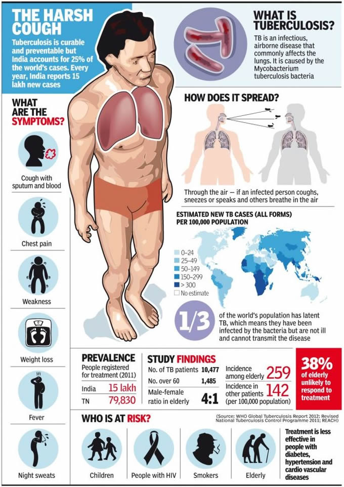
Tuberculosis infographic

Causes and symptoms

Transmission

Tuberculosis spreads by droplet infection, in which a person breathes in the bacilli released into the air when a TB patient exhales, coughs, or sneezes.

However, TB is not considered highly contagious compared to other infectious diseases. Only about one in three people who have close contact with a TB patient, and fewer than 15% of more remote contacts, are likely to become infected.

Unlike many other infections, TB is not passed on by contact with a patient’s clothing, bed linens, or dishes and cooking utensils. Yet if a woman is pregnant, her fetus may contract TB through blood or by inhaling or swallowing the bacilli present in the amniotic fluid.

Once inhaled, water in the droplets evaporates and the tubercle bacilli may reach the small breathing sacs in the lungs (the alveoli), then spread through the lymph vessels to nearby lymph nodes. Sometimes the bacilli move through blood vessels to distant organs.

At this point they may either remain alive but inactive (quiescent), or they may cause active disease. The likelihood of acquiring the disease increases with the concentration of bacilli in the air, and the seriousness of the disease is determined by the number of bacteria with which a patient is infected.

Ninety percent of patients who harbor M. tuberculosis do not develop symptoms or physical evidence of the disease, and their x rays remain negative. They are not contagious; however, these individuals may get sick at a later date and then pass on TB to others.

Though it is impossible to predict whether a person’s disease will become active, researchers surmise that more than 90% of cases of active tuberculosis come from this pool of people. An estimated 5% of infected persons get sick within 12-24 months of being infected.

Another 5% heal initially but, after years or decades, develop active tuberculosis. This form of the disease is called reactivation TB, or post-primary disease. On rare occasions a previously infected person gets sick again after a second exposure to the tubercle bacillus.

Pulmonary tuberculosis

Pulmonary tuberculosis is TB that affects the lungs, and represents about 85% of new cases diagnosed. It usually presents with a cough, which may or may not produce sputum. In time, more sputum is produced that is streaked with blood.

The cough may be present for weeks or months and may be accompanied by chest pain and shortness of breath. Persons with pulmonary TB often run a low-grade fever and suffer from night-sweats. The patient often loses interest in food and may lose weight.

If the infection allows air to escape from the lungs into the chest cavity (pneumothorax) or if fluid collects in the pleural space (pleural effusion), the patient may have difficulty breathing. The TB bacilli may travel from the lungs to lymph nodes in the sides and back of the neck. Infection in these areas can break through the skin and discharge pus.

Extrapulmonary tuberculosis

Although the lungs are the major site of damage caused by tuberculosis, many other organs and tissues in the body may be affected. About 15% of newly diagnosed cases of TB are extrapulmonary, with a higher proportion of these being HIV-infected persons. The usual progression of the disease is to begin in the lungs and spread to locations outside the lungs (extrapulmonary sites).

In some cases, however, the first sign of disease appears outside the lungs. The many tissues or organs that tuberculosis may affect include:
  • Bones. TB is particularly likely to attack the spine and the ends of the long bones.
  • Kidneys. Along with the bones, the kidneys are probably the most common site of extrapulmonary TB. There may, however, be few symptoms even though part of a kidney is destroyed.
  • Female reproductive organs. The ovaries in women may be infected; TB can spread from them to the peritoneum, which is the membrane lining the abdominal cavity.
  • Abdominal cavity. Tuberculous peritonitis may cause pain ranging from the mild discomfort of stomach cramps to intense pain that may mimic the symptoms of appendicitis.
  • Joints. Tubercular infection of joints causes a form of arthritis that most often affects the hips and knees.
  • Meninges. The meninges are tissues that cover the brain and the spinal cord. Infection of the meninges by the TB bacillus causes tuberculous meningitis, a condition that is most common in young children and the elderly. It is extremely dangerous. Patients develop headaches, become drowsy, and eventually comatose. Permanent brain damage can result without prompt treatment.
  • Skin, intestines, adrenal glands, and blood vessels. All these parts of the body can be infected by M. tuberculosis. Infection of the wall of the body’s main artery (the aorta), can cause it to rupture with catastrophic results. Tuberculous pericarditis occurs when the membrane surrounding the heart (the pericardium) is infected and fills up with fluid that interferes with the heart’s ability to pump blood.
  • Miliary tuberculosis. Miliary TB is a life-threatening condition that occurs when large numbers of tubercle bacilli spread throughout the body. Huge numbers of tiny tubercular lesions develop that cause marked weakness and weight loss, severe anemia, and gradual wasting of the body.

Diagnosis

TB is diagnosed through laboratory test results. The standard test for tuberculosis infection, the tuberculin skin test, detects the presence of infection, not of active TB. Skin testing has been done for more than 100 years. In this process, tuberculin is an extract prepared from cultures of M. tuberculosis.

It contains substances belonging to the bacillus (antigens) to which an infected person has been sensitized. When tuberculin is injected into the skin of an infected person, the area around the injection becomes hard, swollen, and red within one to three days.

Today skin tests utilize a substance called purified protein derivative (PPD) that has a standard chemical composition and is therefore a good measure of the presence of tubercular infection.

The PPD test, also called the Mantoux test, is not always 100% accurate; it can produce false positive as well as false negative results. The test may indicate that some people who have a skin reaction are not infected (false positive) and that some who do not react are in fact infected (false negative).

The PPD test is, however, useful as a screener and can be used on people who have had a suspicious chest x ray, on those who have had close contact with a TB patient, and persons who come from a country where TB is common.

Because of the multiple and varied symptoms of TB, diagnosis on the basis of external symptoms is not always possible. TB is often discovered by an aneh chest x ray or other test result rather than by a claim of physical discomfort by the patient. After an irregular x ray, a PPD test is always done to show whether the patient has been infected.

To verify the test results, the physician obtains a sample of sputum or a tissue sample (biopsy) for culture. In cases where other areas of the body might be infected, such as the kidney or the brain, body fluids other than sputum (urine or spinal fluid, for example) can be used for culture.

One important new advance in the diagnosis of TB is the use of molecular techniques to speed the diagnostic process as well as improve its accuracy. As of late 2002, four molecular techniques are increasingly used in laboratories around the world.

They include polymerase chain reaction to detect mycobacterial DNA in patient specimens; nucleic acid probes to identify mycobacteria in culture; restriction fragment length polymorphism analysis to compare different strains of TB for epidemiological studies; and genetic-based susceptibility testing to identify drug-resistant strains of mycobacteria.

Treatment

Because of the nature of tuberculosis, the disease should never be treated by alternative methods alone. Alternative treatments can help support healing, but treatment of TB must include drugs and will require the care of a physician. Any alternative treatments should be discussed with a medical practitioner before they are applied.

Supportive treatments include:
  • Diet. Nutritionists recommend a whole food diet including raw foods, fluids, and particularly pears and pear products (pear juice, pear sauce), since pears may help heal the lungs. Other helpful foods include fenugreek, alfalfa sprouts, garlic, pomegranate, and yogurt or kefir. Four tablespoons of pureed steamed asparagus at breakfast and dinner taken for a few months may also be helpful.
  • Nutritional therapy. Nutritionists may recommend one or many of the following vitamins and minerals: vitamin A at 300,000 IU for the first three days, 200,000 IU for the next two days, then 50,000 IU for several weeks; beta-carotene at 25,000-50,000 IU; vitamin E at up to 1,000 IU daily unless the patient is a premenopausal woman with premenstrual symptoms; lipotrophic formula (one daily); deglycerolized licorice; citrus seed extract; vitamin C; lung glandular; essential fatty acids; vitamin B complex; multiminerals; and zinc.
  • Herb therapy may use the tinctures of echinacea, elecampane, and mullein taken three times per day, along with three garlic capsules three times per day.
  • Hydrotherapy may be used up to five times weekly. Dr. Benedict Lust, the founder of naturopathy, supposedly cured himself of tuberculosis by using hydrotherapy.
  • Juice therapy. Raw potato juice, may be taken three times daily with equal parts of carrot juice plus one teaspoon of olive or almond oil, one teaspoon of honey, beaten until it foams. Before using the potato juice, starch should be allowed to settle from the juice.
  • Topical treatment may use eucalyptus oil packs, grape packs or grain alcohol packs.

Professional practitioners may also treat tuberculosis using cell therapy, magnetic field therapy, or traditional Chinese medicine. Fasting may be undertaken, but only with a doctor’s supervision.

Allopathic treatment

Drug therapy

Five drugs are most commonly used today to treat tuberculosis: isoniazid (INH), rifampin, pyrazinamide, streptomycin, and ethambutol. Of the five medications, INH is the most frequently used drug for both treatment and prevention. The first three drugs may be given in the same capsule to minimize and treat active TB the number of pills in the dosage.

As of 1998, many patients are given INH and rifampin together for six months, with pyrazinamide added for the first two months. Hospitalization is rarely necessary because many patients are no longer infectious after about two weeks of combination treatment. A physician must monitor side effects and conduct monthly sputum tests.

In 2002, the Centers for Disease Control (CDC) worked with medical organizations to release new guidelines that better individualize the drug regimens received by TB patients depending on their disease symptoms and severity. Many can now receive once-weekly doses of rifapentine in the continuation phase of treatment.

The first large scale trial of a new agent to treat TB began in 2002. The promising new drug, called moxifloxacin, may mean a shorter treatment course for TB sufferers in the near future.

It will also be tested in combination with rifapentine, and researchers believe that using the drugs together will mean a less frequent dosing schedule for patients.

Drug resistance has become a dilema in treating TB. When patients do not take medication properly or for long enough periods of time, the TB organisms may become drug resistant. This makes the patient vulnerable to further infection and allows the TB organism to develop resistance.

Surgery

Surgical treatment of TB may be used if medications are ineffective. There are three surgical treatments for pulmonary TB: pneumothorax, in which air is introduced into the chest to collapse the lung; thoracoplasty, in which one or more ribs are removed; and removal of a diseased lung, in whole or in part. It is possible for patients to survive with one healthy lung.

Expected results

The prognosis for recovery from TB is good for most patients, if the disease is diagnosed early and given prompt treatment with appropriate medications on a long-term regimen.

According to a 2002 Johns Hopkins study, most patients in the United States who die of TB are older—average age 62— and suffer from such underlying diseases as diabetes and kidney failure.

Modern surgical methods are usually effective when necessary. Miliary tuberculosis is still fatal in many cases but is rarely seen today in developed countries.

Even in cases in which the bacillus proves resistant to all of the commonly used medications, other seldom-used drugs may be tried because the tubercle bacilli have not yet developed resistance to them.

Prevention

Vaccination is widely used as a prevention measure for TB. A vaccine called BCG (Bacillus Calmette-Guérin, named after its French developers) is made from a weakened mycobacterium that infects cattle.

Vaccination with BCG does not prevent infection, but it does strengthen the immune system of first-time TB patients. As a result, serious complications are less likely to develop. BCG is used more widely in developing countries than in the United States.

Though the vaccine has been proven beneficial and fairly safe, its use is still controversial. It is not clear whether the vaccine’s effectiveness depends on the population in which it is used or on variations in its formulation. Recently, efforts have been focused on developing a new vaccine.

Generally, prevention focuses on the prevention of transmission, skin-testing high-risk persons and providing preventive drug therapy to people at risk.

Measures such as avoidance of overcrowded and unsanitary conditions are necessary aspects of prevention. Hospital emergency rooms and similar locations can be treated with ultraviolet light, which has an antibacterial effect.

INH is also given to prevent TB, and decreases the incidence of TB by about 60% over the life of the patient. INH is effective when taken daily for 6 to 12 months by people in high-risk categories who are under 35 years of age.

About 1% of patients in preventive treatment develop toxicity. Because INH carries the risk of side effects (liver inflammation, nerve damage, changes in mood and behavior), it is important for its use to be monitored and to give it only to persons at special risk.

Unfortunately, failure of TB patients to complete the full course of their drugs adds to TB incidence and encourages development of drug-resistant strains of the disease.

As scientists try to develop drugs that require shorter courses, physicians must work with patients to encourage compliance with their treatments. Even if symptoms go away, patients often have to continue their drug treatment for six months to be sure to stop the spread of their TB infection to others.

Tibetan
Tibetan Medecine - chakras and energy channels

Tibetan medicine differs from allopathic medicine in that it has no concept of illness as such, but rather the concept is of disharmony of the organism. Accordingly, this system of medicine, like many alternative therapies, seeks to achieve a harmony of the self.

Medicine is one of five branches of Tibetan science, and is known to the Tibetans as gSoba Rig-pa—the science of healing.

The Tibetan pharmacopoeia utilizes many different elements in the treatment of disease, such as trees, rocks, resins, soil, precious metals, sap, and so on, but like Chinese medicine, to which it is related, it mainly relies on herbs for treatment.

TibetanTibetan

Origins

Tibetan medicine, like its relative Chinese medicine, is an ancient art that has become associated with many legends and is surrounded by a cloud of mysticism. Although Tibetan culture is more recent, Tibetan medical practices can be traced back over 2,500 years. It is now practiced in secret or by those in exile since Communist rule has suppressed it in its country of origin.

The treatise of Tibetan medicine, which can be described as a manual compiled over thousands of years, is called the Chzud-shi. In addition to the medical theory, this manual also incorporates the Tibetan pharmacopoeia.

Tibetan
Medicine Buddha with healing herbs

Benefits

Tibetan medicine has been particularly successful at treating chronic conditions such as rheumatism, arthritis, ulcers, digestive problems, asthma, hepatitis, eczema, liver disorders, sinus problems, emotional disorders and nervous system problems. Like many alternative therapies, it is a holistic therapy that treats the whole person and encourages a healthy way of life that will promote well-being at all levels.

Description

Harmony and the balance of all aspects of the human organism are the concepts that form the basis for Tibetan medicine. The three elements that must be kept in harmony are known collectively as the Nyipa sum, and they are rLung, mKhris-pa, and Bad-kan.

It is said that the Tibetan words describing their medicine are very difficult to translate, rather an explanation of the meaning is attempted. Desire, hatred, and delusion are considered to be very harmful influences affecting this harmony, and illustrate the close connection between the Tibetan medical art and Buddhist teachings.

rLung is considered to be a “subtle flow of energy” that is most closely connected with the “air” element. However, since all five elements; earth, water, fire, air and space, in addition to the concepts of heat and cold play a complex role in the health of the individual, this is no simple matter. All elements and aspects are held to be interdependent.

Types of rLung:
  • Srog-’dzin (life-grasping rLung). Located in the brain, this energy governs swallowing of food, breathing, spitting, sneezing, and the clearing and steadying of the mind.
  • Gyen-rgyu (rLung moving upwards). Located in the chest, it governs speech, physical vigor, general health, and appearance of skin.
  • Khyab-byed (all pervading rLung). Located in the stomach, it governs digestion, metabolism, and the seven physical sustainers referred to as lus-zung dhun.
  • Thur-sel (downward cleansing rLung). Located in the rectum, it governs the elimination of waste products and reproductive fluids in addition to the birth process (for women).

Types of mKhris-pa:
  • mKhris-pa is the heat of human nature, related to fire, described as oily, sharp, hot, light, pungent and moist. Its major function is to balance body temperatures. It governs hunger and thirst, and regulates skin condition. There are five types of mKhris-pa:
  • Ju-byed. This is located between the stomach and the intestine. Governs digestion and assimilation, providing heat and energy.
  • SGrub-byed. Located in the heart. Responsible for anger, aggression, and hatred, and is considered to lead to desire, achievement, and ambition.
  • mDangs-sgur. Located in the liver, it is responsible for maintaining and promoting color and essential components of blood.
  • mThong-byed. Located in the eye, it governs vision.
  • mDog-sel. Located in the skin, it governs skin appearance and texture.

Types of Bad-kan:
  • rTen-byed (supporting Bad-kan). Located in the chest, plays a supporting role to the other four types of Bad-kan.
  • Myag-byed (mixing Bad-kan). Located in the upper half of the body. Mixes nutrients (liquids and solids).
  • Myong-byed (experiencing Bad-kan). Located in the tongue, governs experience of taste.
  • Tsim-byed (satisfaction Bad-kan). Located in the head. Governs the five senses and responsible for heightening their power.
  • Byor-byed (joining Bad-kan). Located in the joints, it is considered responsible for their flexibility.
Tibetan
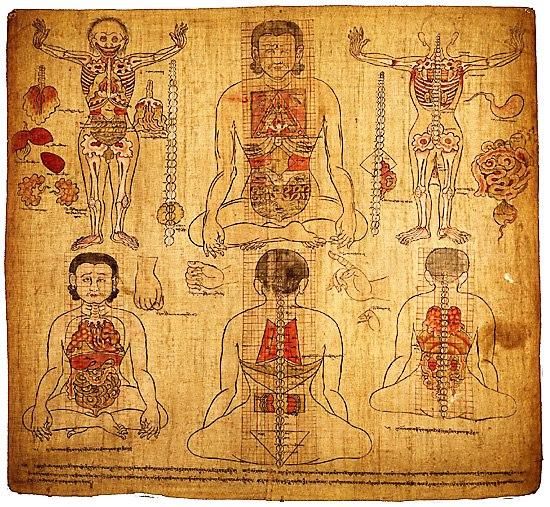
Old Tibetan Medicine painting of anatomy

When these components of Nyipa sum are balanced, the seven bodily sustainers will also be in harmony. They are essential nutrients, blood, muscle tissue, fat, bone, marrow, and reproductive fluids.

Diagnosis

A practitioner of Tibetan medicine will employ several diagnostic tools. Chief of these is a very complicated system of pulse reading, which involves 13 different positions with a possibility of over 300 different readings.

This is similar to traditional Chinese medicine and Ayurvedic medicine. The pulse is likened to a messenger between doctor and patient. For this diagnosis to be effective, it is necessary for the patient to be rested and relaxed.

Another tool of diagnosis is observation, which consists of urinalysis and examining the tongue. To examine the urine, a physician will assess the color, vapor, odor, bubbles, sediments, and albumin content. The color of urine is determined by food and drink, the seasons, and whatever diseases the patient suffers from.

The selesai tool of diagnosis is questioning. The physician will ask specific questions of his patient, and will include such questions as how and when the illness started, where pain is felt, and if the condition is affected by foods eaten.

Treatment

Treatment is divided into four categories, which are dietary advice, lifestyle recommendations, the prescription of medicine, and if necessary, surgical procedures, according to the type of patient.

Treatment proceeds in this order according to the seriousness of the disorder. For example, minor problems are considered to need merely a reassessment of dietary habits, but only in the most serious cases will surgery be considered.

Preparations

A Tibetan physician prescribes medicines and recommends surgery as a last resort. When it is necessary, the prescription is likely to be made up from certain herbs in the form of a decoction, powder, or pills. The prescription will be made up at one of the branches of the Tibetan Medical Institute specifically for each patient.

Precautions

The qualifications of any Tibetan physician should be checked before treatment proceeds.

Side effects

As a natural therapy, Tibetan medicine, if administered correctly, is not known to be associated with any side effects. According to the primary Tibetan medical treatise, one of the criteria for medical prescriptions is that they should be absolutely harmless.

Research and general acceptance

The Tibetan system of medicine has roots in medical practices over 2,500 years old, so it can be considered well researched. Despite the Communist crackdown in Tibet, and the oppression and persecution of their physicians, the Tibetan people still prefer to seek the advice of a traditional physician rather than take advantage of “new” systems of medicine.

In 1994, the Natural Medicine Research Unit, (NMRU) of Hadassah University Hospital in Jerusalem began a double-blind randomized clinical trial of Tibetan herbal formulas which had been on sale in Switzerland for more than seventeen years. Previous trials had already demonstrated the harmlessness of these formulas. The aim of the unit is to compile a database of Tibetan formulas.

Tibetan
The father of Tibetan Medicine, Yuthok sitting in mandala

Training and certification

The headquarters of the main Tibetan medical institute is now in Dharamsala in northern India. Tibetan medicines are also manufactured there. The minimum period of training for a Tibetan physician is seven years.

The first five years mainly consist of theoretical training, and for the sixth and seventh years, medical students are sent for a period of practical training under a senior physician at one of the Institute’s branches, of which there are over 30 in India and Nepal.

Trigger
Trigger point therapy

Trigger point therapy is a bodywork technique that involves the application of pressure to tender muscle tissue in order to relieve pain and dysfunction in other parts of the body. It may also be called myofascial (myo meaning muscle, fascial meaning connective tissue) trigger point therapy.

Trigger point therapy is sometimes regarded as one of a group of treatment aproaches called neuromuscular therapy or NMT. Myotherapy, developed by Bonnie PruddenTrigger, is a related type of trigger point therapy.

Origins

Trigger point therapy was developed by pain experienced in one part of the body is actually caused by an injury or dysfunction in another part of the body.

TriggerTrigger

Ultimately, she mapped what she termed the body’s trigger points and the manner in which pain radiates to the rest of the body. Travell’s work came to national attention when she treated President John F. Kennedy for his back pain.

Trigger points are thought to result from a variety of causes, including birth trauma, hypoglycemia, vitamin B6 deficiency, food allergies, traumatic injuries, poor posture, skeletal asymmetry, overexertion, or such diseases of the digestive tract as ulcers and irritable bowel syndrome. During times of physical or emotional stress, the points cause muscles to spasm.

Travell’s therapy called for the injection of saline (a salt solution) and procaine (also known as Novocaine, an anesthetic) into the trigger point. Although beneficial in the relief of pain, the injections are a painful procedure for some people.

In the 1970s, Bonnie Prudden, a physical fitness and exercise therapist, found that applying sustained pressure to a trigger point also relieved pain. Prudden developed her techniques over a number of years and called the treatments myotherapy. Myotherapy is beneficial to patients who find that trigger point injections are too painful.

Benefits

Trigger point therapy is said to interrupt the neural signals that cause both the trigger point and the pain. The object is to eliminate pain and to reeducate the muscles into pain-free habits.

In this manner, the swelling and stiffness of neuromuscular pain is reduced, range of motion is increased, and flexibility and coordination are improved. The therapy can also relieve tension and improve circulation.

The list of conditions that benefit from trigger point therapy include arthritis; carpal tunnel syndrome; chronic pain in the back, knees, and shoulders; headaches; menstrual cramps; multiple sclerosis; muscle spasms, tension, and weakness; postoperative pain; sciatica; temporomandibular joint syndrome (TMJ); tendinitis; and whiplash injuries.

Description

Typically, a health care professional refers a patient to a trigger point therapist. The therapist will take a history of injuries suffered, occupations held, and sports played. He or she will ask the individual to describe the pain and its location in detail.

The therapist will then probe the area of the coordinating trigger point. An injection of lidocaine, saline, or other medicines, or probing with a dry needle, may be done. In myotherapy, once the point is found, the therapist will apply sustained pressure using the fingers, knuckles, or elbows for several seconds.

Pain relief is often experienced immediately. Following the injection or pressure treatment, the therapist will then gently stretch the muscles of the trigger point. Finally, a series of exercises is taught to the individual to reeducate the muscles and to prevent the pain from returning.

Workbooks are now available to help patients maximize the benefits of trigger point therapy through self-treatment at home.

Trigger
trigger point referral pain pattern for the mid back

Preparation

Persons should consult a health care professional before beginning trigger point therapy to insure that the pain is not caused by fracture or disease. In fact, a certified trigger point therapist will not provide services to someone who is not referred by a health care professional.

The therapy is usually conducted on a padded table or treatment chair. The individual should wear comfortable loose-fitting clothing. An ongoing, honest interaction with the therapist will facilitate the sessions.

Treatment sessions can last 30 minutes to an hour. The range of cost is approximately $45–60 per session. Acute pain can be relieved in as little as one session. Chronic pain may require numerous treatments.

Precautions

Persons with infectious diseases, open sores, or recent injuries should wait until they have recovered before beginning trigger point therapy.

Persons taking anticoagulant prescription drugs may experience bruising after trigger point therapy.

Research and general acceptance

Research into the effects of trigger point therapy is sketchy, although the growing acceptance of acupuncture within the mainstream medical community has led to a few recent published studies of trigger point therapy.

Interest in trigger point therapy is growing in Europe and Asia as well as in the United States; one recent study by a group of Japanese researchers reported that trigger point therapy was superior to standard allopathic drugs in relieving the pain of renal colic.

The American Academy of Pain Management (AAPM) reports that studies of trigger point therapy on back pain and headaches have been conducted on groups of fewer than 10 people. The AAPM does, however, recognize trigger point therapy as a valid approach to the management and relief of pain.

In the traditional medical community, trigger point therapy is viewed as a complement to treatment. Patients are referred by a variety of health professionals including psychiatrists, orthopedic surgeons, and anesthesiologists.

Trepanation
Trepanation

Trepanation is a surgical procedure in which a circular piece of bone is removed from the skull by a special saw-like instrument called a trephine or trepan. The operation is also known as trephination or trephining. The English word “trepan” comes from the Greek word trypanon, which means “auger” or “drill.”

In standard medical practice, trepanation is occasionally performed by a neurosurgeon in order to relieve pressure on the brain caused by trauma, or to remove a blood clot from brain tissue.

In recent years, however, trepanation has been touted by a small group of alternative practitioners as a way to expand one’s consciousness through the increase of blood flow to the brain and opening the “third eye,” also known as the inner eye or eye of the mind.

TrepanationTrepanation

Practitioners of kundalini yoga refer to the opening of the third eye, located in the middle of the forehead, as entry into a new and completely different dimension of reality.

Trepanation is the oldest surgical procedure known to humans; skulls of Cro-Magnon people estimated to be 40,000 years old have been discovered with circular holes as large as 2 in in diameter. The Incas of Peru are known to have performed trepanation as early as 2000 B.C.

It is thought that these operations were performed to treat people suffering from psychotic disorders, epilepsy, or chronic migraine headaches by allowing demons to escape through the hole in the skull.

The oldest written reference to trepanation comes from Hippocrates (c. 400 B.C.), whose descriptions of head injuries refer to it as a necessary treatment for skull fractures with bone fragments pushed inward and compressing the brain.

Celsus and Galen refer to Roman surgeons of the first century A.D. as performing trepanations with implements resembling carpenters’ drills. Trephines were refined in various ways through the Middle Ages, the Renaissance, and the eighteenth and nineteenth centuries.

It should be emphasized that trepanations were done by ancient, medieval, and early modern physicians to relieve pressure on brain tissue—not to perform surgery on the brain itself.

Care was taken not to penetrate the dura mater, which is the outermost of the three meninges or membranes that lie beneath the skull and form a protective cover for the brain and spinal cord. Historians of medicine estimate, however, that as many as 40 percent of patients died from infections following the procedure rather than from the surgery itself.

Contemporary interest in trepanation as a path to expanded consciousness goes back only to the 1960s.

Bart Huges, a Dutchman who was expelled from medical school in the early 1960s for failing his examinations and using marijuana, is generally considered the founder of alternative trepanation. Huges developed a theory that he called brainbloodvolume while he was smoking marijuana at a party on the island of Ibiza.

He noticed another guest standing on his head to increase the intoxicating effects of the drug. Huges concluded that the expansion of consciousness associated with hallucinogens results from an increased volume of blood in the brain.

Trepanation
Japanese Anatomical Illustrations of trepanation

He reasoned that the removal of a piece of the skull would allow an even larger amount of blood to enter the brain, speeding up the delivery of oxygen and glucose to the brain cells as well as the removal of toxins.

Huges had also learned in medical school that infants are born with soft spots in the skull known as fontanelles, which are membrane-covered areas where the bone has not yet completely formed. He concluded that trepanation would help to return an adult’s consciousness to the intense imagination and vivid dreams of a child.

Huges—who never obtained a medical degree—managed to convert several individuals to his brainbloodvolume theory—among them Peter Halvorson, who underwent trepanation and credits it with curing his depression, increasing his energy level, and giving him a permanent drug-free high. As of 2004, Halvorson is the head of the International Trepanation Advocacy Group (ITAG), headquartered in Wernersville, Pennsylvania.

The ITAG web site includes accounts of a pilot study of six volunteers who were trepanned in June 2002 as well as personal testimonials from others who have undergone the procedure.

Benefits

According to the testimonials collected by Halvorson, trepanation confers the following benefits:
  • relief from anxiety, depression, and other mood disorders
  • feelings of freedom and serenity
  • a richer emotional life
  • greater ability to recall dreams on awaking
  • decrease in frequency and severity of chronic headaches
  • higher energy levels

Other people who have undergone trepanation, however, maintain that these benefits are only temporary and may be due to the placebo effect.

A man who performed trepanation on himself in 2000 reported to an interviewer from an online body modification journal that he had “come to the frustrating conclusion [four weeks after the procedure] that the trepanation has had no lasting effect ... Trepanation has no more physiological effect than any other syok ... it does not do what many hope it will.”

Description

Surgical trepanation

A standard trepanation—most commonly done to relieve pressure on the brain when a portion of the skull has been pushed inward—is performed with the patient under general anesthesia under sterile conditions.

The neurosurgeon cuts the scalp over the injured area, pulls back a flap of skin, and bores a hole in the underlying skull with a trephine. After the depressed bone has been removed together with any blood clots that have formed, the surgeon carefully cleanses the area and closes the incision.

Alternative trepanation

Some alternative trepanations have been performed by people on themselves, with friends to assist with the procedure. In the early 1980s, several people in England performed the entire operation on themselves, with others present to help only if an emergency arose.

The reason for this stipulation was to protect the others in the room from criminal prosecution for performing surgery without credentials. The trepanner typically shaved his or her head and injected a local anesthetic. He or she then made an incision in the scalp over the area to be trepanned.

Next, a hole between 1/4 and 1/2 in in diameter was cut in the skull with a foot-powered dental drill. The trepanner then removed the piece of skull, cleaned the incision, and bandaged it. The scalp gradually grew back over the hole, leaving only a small permanent indentation.

More recently, however, trepanners have allowed others to assist with the operation; the man who was interviewed for the online journal had three friends who covered the walls of his room with plastic sheeting, did part of the drilling, and rinsed out the incision from time to time with sterile saline solution. He reported that the entire procedure took about 3-1/2 hours.

The participants in the ITAG pilot study, however, went to a clinic in Monterrey, Mexico, for their trepanations. The ITAG web site states plainly that “Self-trepanation today is a very selfish act. It opens the door for no one and you’d always have to keep it a secret. The public mind can’t handle this. You’d be labeled ‘insane.’”

According to the ITAG web site, the surgeon who presently performs the procedures for Halvorson’s groups was trained in Texas and is board-certified in four countries (France, Spain, Mexico, and the United States). The trepanations take about 35 minutes to complete. The cost of the operation is $2400–$3600, not including travel and hotel fees.

Preparations

People who have performed trepanations on themselves have prepared by assembling the needed equipment and setting aside a room in their house to serve as the operating room.

ITAG requires persons interested in trepanation (who must be 18 or older) to go through a period of mental preparation known as engramming, which Halvorson defines idiosyncratically as “becom[ing] thoroughly acquainted with the terminology of conscious expansion.”

In addition, the volunteers must sign an informed consent form and a protocol that indicates that they understand the procedure is considered experimental. They are given MRIs before and after the trepanation.

Precautions

Mainstream medical professionals uniformly warn against alternative trepanation because it is an extremely risky procedure—particularly if done by amateurs—with no certain or permanent benefits.

Because scalp incisions bleed profusely, people who attempt to trepan themselves are likely to find that the flow of blood obscures their field of vision, thus increasing the risk of self-injury.

Side effects

The potential side effects of alternative trepanation are severe, even life-threatening; they include permanent injury or death from infections, stroke, direct damage to brain tissue, generalized encephalitis, epilepsy, or brain abscesses.

Research and general acceptance

Trepanation is not accepted as an alternative therapy by any mainstream physicians or surgeons in the United States or Canada. In addition to the dangers of the procedure itself, neurosurgeons who have studied the claims made for trepanation say that Huges’ brainbloodvolume theory is anatomically impossible.

Contact Form

Name

Email *

Message *

Powered by Blogger.